 Imaging deep structures of the body using high-frequency sound waves.
The visualization of deep structures of the body through the recording of high-frequency sound waves directed into tissues, ultrasound device.
The procedure of passing ultrasound waves through the body and recording echoes which show details of internal organs.
Process by which the reflection of high-frequency sound waves is used to develop an image (sonogram) of a stracture; used in medicine to study fetal growth and detect abnormalities, and to study the heart and many other organs.
The measurement of interior body structures by means of ultrasonic waves, with special computerized equipment that can translate the reflection of the sound waves into images.
The use of ultrasound to produce pictures of structures within the body.
The use of ultrasound to produce images of structures in the body that can be viewed on a television screen and transferred to photographic film.
The use of ultrasound to produce an image or photograph of an organ or tissue. Ultrasonic echoes are recorded as they return from reflecting or refracting tissues of different densities.
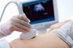
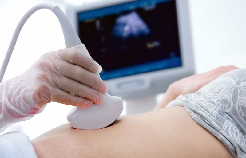
A diagnostic examination that utilizes sound waves to assess blood flow involves the application of gel on a handheld transducer, which is then pressed against the patient’s body. The resulting images are displayed on a monitor for analysis and interpretation.