
 High-intensity sound waves used to create an image of internal body structures.
Very high frequency sound waves which can be reflected off internal body parts or off a fetus in the womb to create images for medical examination.
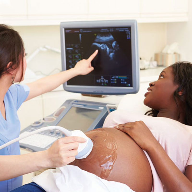
Pulses of high-frequency sound waves used in medicine to create a moving image, or sonogram, of body tissues on a television monitor. During pregnancy, ultrasound is often employed to check the progress of fetal development, identify multiple pregnancy, learn the fetus’s sex, identify the position of the fetus and placenta (should pre-birth intervention be necessary), and check for abnormalities, such as anencephaly or ectopic pregnancy, especially if the fetus size is inappropriate for the presumed stage of pregnancy or if family history warrants. Carrying little or no risk to mother or fetus, ultrasound scanning is also used as a guide to physicians in some other genetic screening procedures, such as amniocentesis and chorionic villus sampling. In newborns, it is sometimes used to peer through gaps in the skull, called fontanelles, for brain abnormalities, such as hydrocephalus or BRAIN tumors. In patients of all ages, ultrasound examination can help physicians check for abnormalities in internal tissues, such as those of the liver and gall bladder; special ultrasound examination of the heart is called echocardiography. Ultrasound can also help physicians identify possible structural causes of infertility.
Sound with a pitch above human hearing (above 20,000 Hz). Ultrasound is used in an imaging technique to visualize internal structures by recording the reflection of the sound waves by the tissues. Ultrasound is also used in some forms of therapy, such as the liquidizing of cataracts and their removal by suction, a process called “phacoemulsufication.”
Sound waves at very high frequencies used in the technique of ultrasonography to aid diagnosis.
Noninvasive procedure that generates images via computer analysis to assess blood-flow velocity, direction, and occlusions; uses a transducer, which is placed over the blood vessel, and a computer, which analyzes the echoes for aberrations.
High-frequency sound waves used to project images of various parts of the body. A transducer applied to the skin produces the ultrasound and catches the response, which is displayed on an oscilloscope. Often used to assess fetal development at various stages of pregnancy.
Sound waves of extremely high frequency (above 20,000 Hz), inaudible to the human ear. Ultrasound can be used to examine the structure of the inside of the body, in the same way that X-rays can be used to build up pictures but with the advantages that the patient is not submitted to potentially harmful radiation and that structures not opaque to X-rays can be seen. The vibratory effect of these sound waves can also be used in the treatment of various disorders of deep tissues, and even to break up stones in the kidney or elsewhere.
Ultrasound, or ultrasonic, waves comprise very high- frequency sound waves above 20,000 Hz that the human ear cannot hear. Ultrasound is widely used for diagnosis and also for some treatments. In obstetrics, ultrasound can assess the stage of pregnancy and detect abnormalities in the fetus. It is a valuable adjunct in the investigation of diseases in the bladder, kidneys, liver, ovaries, pancreas and brain (for more information on these organs and their diseases, see under separate entries); it also detects thromboses (clots) in blood vessels and enables their extent to be assessed. A noninvasive technique that does not need ionising radiation, ultrasound is quick, versatile and relatively inexpensive, with scans being done in any plane of the body. There is little danger to the patient or operator: unlike, for example, x-rays, ultrasound investigations can be repeated as needed. A contrast medium is not required. Its reliability is dependent upon the skill of the operator.
Inaudible sound in the frequency range of approx. 20,000 to 10 billion (109) cycles/sec. Ultrasound has different velocities that differ in density and elasticity from one kind of tissue to the next. This property permits the use of ultrasound in outlining the shape of various tissues and organs in the body. In obstetrics, for example, identifying the size and position of the fetus, placenta, and umbilical cord enables estimation of gestational age, detects some fetal anomalies and fetal death, and facilitates other diagnostic procedures, such as amniocentesis. In physical therapy, the thermal effects of ultrasound are used to treat musculoskeletal injuries by warming tissue, increasing tissue extensibility, and improving local blood flow. Ultrasound is used to facilitate movement of certain medications (e.g., pain relievers) into tissue (phonophoresis). Ultrasound is also used with electric current for muscular stimulation. The diagnostic and therapeutic uses of ultrasound require special equipment.
Sound waves that exceed the frequency of 20,000 cycles per second or 20,000 Hertz (Hz), surpassing the audible range of human hearing.
A medical modality that employs high-frequency sound waves to facilitate diagnosis and treatment by generating visual representations of internal organs is known as ultrasound imaging.
Sound waves possessing a frequency surpassing the upper threshold of human hearing, which is over 20,000 hertz (cycles per second), are termed ultrasound. In the medical field, ultrasound is employed to generate images of bodily structures, facilitating diagnosis, or as a therapeutic tool to enhance healing and eliminate anomalies like kidney stones. The frequency range utilized for these medical applications usually falls between 1 to 15 million hertz.