 Hearing and balance organ, including three general structures; the outer (external) ear; the middle ear, including the eardrum cavity and the three tiny bones that transmit the hearing vibrations; and the inner (internal) ear, including the main organ of hearing (the organ of Corti) and the balance mechanism.
The organ for hearing and balance. The structures of the ear are in three parts: the outer ear, the middle ear, and the inner ear. Most of these structures lie inside the skull. The outer ear receives sound waves from the environment and channels them to the middle ear, the middle ear transmits them to the inner ear, and the inner ear converts the sound waves into impulses that are sent to the brain.
The sense organ concerned with hearing and balance. Sound waves, transmitted from the outside into the external auditory meatus, cause the eardrum (tympanic membrane) to vibrate. The small bones (ossicles) of the middle ear, the malleus, incus, and stapes, transmit the sound vibrations to the fenestra ovalis, which leads to the inner ear. Inside the cochlea the sound vibrations are converted into nerve impulses. Vibrations emerging from the cochlea could cause pressure to build up inside the ear, but this is released through the Eustachian tube. The ‘semicircular canals, saccule, and utricle (also in the inner ear) are all concerned with balance.
The ear is concerned with hearing and the senses of equilibration and of motion. It consists of three parts: (1) the external ear, consisting of the auricle on the surface of the head, and the tube which leads inwards to the drum; (2) the middle ear, separated from the former by the tympanic membrane or drum, and from the internal ear by two other membranes, but communicating with the throat by the Eustachian tube; and (3) the internal ear, comprising the complicated labyrinth from which runs the vestibulocochlear nerve into the brain.
The organ of hearing and equilibrium. The ear consists of outer, middle, and inner portions, and is innervated by the eighth cranial nerve.
The sense organ that functions in hearing and balance.
The ear is the intricate organ responsible for both hearing and balance. It comprises three essential parts: the outer ear, the middle ear, and the inner ear. The primary functions of the outer and middle ear revolve around collecting and transmitting sound waves. On the other hand, the inner ear plays a crucial role in analyzing sound waves and houses the mechanism responsible for maintaining the body’s balance.
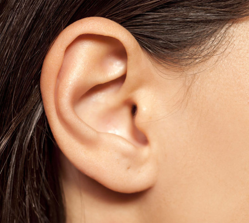
The outer ear consists of two main parts: the pinna, which is the visible outer portion of the ear, made up of skin and cartilage folds, and the ear canal, measuring approximately 2.5 cm in length for adults.
The external section of the ear canal is made up of cartilage and generates earwax, which captures dust and foreign objects. The canal is sealed at its innermost point by the eardrum, a delicate, circular membrane covered with a layer of skin. This eardrum vibrates when exposed to variations in air pressure, which make up sound.
The middle ear serves as a small chamber situated between the eardrum and the inner ear, responsible for transmitting sound to the inner ear through three minuscule interconnected and movable bones called ossicles. The first bone, known as the malleus, is attached to the inner surface of the eardrum. The second bone, the incus, forms a wide joint with the malleus, almost parallel to it, and has a delicate joint with the third bone, the stapes. The base of the stapes fills the oval window, which leads to the inner ear.
The middle ear is isolated from the external environment by the eardrum. Nevertheless, it is not entirely sealed off; there exists a ventilation passage known as the eustachian tube, which extends forward and downward into the back of the nose. Ordinarily, the eustachian tube remains closed, but it opens through muscular contractions when a person yawns or swallows.
The middle ear functions as a transformer, transferring sound vibrations from the thin medium of the air outside to the thicker medium of the fluid within the inner ear.
Deeply nestled within the bones of the skull, the inner ear comprises a complex arrangement of structures. It forms a labyrinth, a winding maze of passages. The foremost section, known as the cochlea, bears a resemblance to a snail’s shell and houses nerve fibers that are responsible for detecting various sound frequencies.
The posterior section of the inner ear holds three semicircular canals, which play a crucial role in maintaining balance. These canals are linked to a space known as the vestibule and house hair cells immersed in fluid. Some of these cells are sensitive to gravity and acceleration, while others respond to the orientation and motion of the head. The information gathered in the inner ear is transmitted to the brain through the vestibulocochlear nerve.
The organ of hearing. The external ear consists of the pinna or ear flap and the external auditory meatus, the tube from the outer ear to the eardrum. The middle ear is the cavity between the eardrum and the bony wall of the inner ear; it contains three small bones—the incus, the malleus, and the stapes. These small bones, which articulate with one another, are attached to the eardrum, or the tympanic membrane, and transmit its vibrations to the inner ear. The inner ear, or labyrinth, consists of the vestibule (a cavity containing a fluid), into the front of which open the cochlea, or spiral canal, containing the essential organ of hearing (the organ of Corti), and the semicircular canals responsible for the sense of balance. Extreme motion upsetting the movements within the semicircular canals causes severe vertigo and the sensations of motion sickness, as seasickness and airsickness.