 Imaging that uses magnetic fields and radio waves to produce high‐quality two‐ or three dimensional images without use of ionizing radiation (X‐rays) or radioactive tracers. During an MRI scan, a large cylindrical magnet creates a magnetic field around the patient through which radio waves are sent. Medical MRI most frequently relies on the relaxation properties of excited hydrogen nuclei in water. The vast quantity of nuclei in a small volume sum to produce a detectable change in a magnetic field, which can be measured from outside the body. When the magnetic field is imposed, each point in space has a unique radiofrequency at which the signal is received and transmitted. Sensors read the frequencies and a computer uses the information to construct an image.
A technique for visualizing anatomical structures. It involves placing patients in a strong magnetic field and then, by use of magnetic gradients and brief radio frequency pulses, determining the resonance characteristics at each point in the area to be studied. Used to detect structural or anatomical abnormalities, such as brain tumor and incipient multiple sclerosis. In psychiatry, neurology, and neurosurgery, it is used to detect structural or anatomical abnormalities. It is better able to differentiate between gray and white matter than is computed tomography (CT).
A diagnostic imaging technique that uses magnetic fields and radio waves to visualize internal structures.
A procedure similar to computed tomography that does not require radiographs; a large magnetic field is applied and creates an image; useful in visualizing the cardiovascular system, brain, and soft tissues.
An imaging technique employing magnetic energy rather than X rays.
A technology allowing the imaging of a body without radiation hazard.
Diagnostic imaging by use of electromagnetic radiation to visualize soft tissues of the body.
A procedure in which a scanning device uses a magnetic field together with radiofrequency energy that allows penetration of bone to visualize soft tissue to determine abnormal conditions of brain and spinal cord soft tissues. Such conditions as edema (swelling of tissues due to excessive fluid accumulation), tumors, and diseases such as multiple sclerosis may be diagnosed with this technique.
A diagnostic imaging technique that uses powerful magnetic fields and radio-frequency waves to produce computer-enhanced, cross-sectional images of internal organs and structures. MRI allows physicians to visualize interior body parts with great clarity and without exposure to the radiation involved in the use of X rays. An MRI is generally used to obtain two-dimensional views of an internal organ or structure, particularly of soft tissues (for example, muscles) that do not show up well on regular X rays. The technique is particularly helpful for visualizing internal areas of the nervous system, including the brain and spinal cord. It may also be used to evaluate injuries to the bones and joints. MRI is often used to monitor response to chemotherapy, radiation therapy, or other treatments for cancer and other diseases.
A type of medical imaging that uses the characteristic behavior of protons when placed in powerful magnetic fields to make images of tissues and organs. Certain atomic nuclei with an odd number of neutrons, protons, or both are subjected to a radiofrequency pulse, causing them to absorb and release energy. The resulting current passes through a radiofrequency receiver and is then transformed into an image. This technique is valuable in providing soft tissue images of the central nervous and musculoskeletal systems. Imaging techniques allow visualization of the vascular system without the use of contrast agents. Agents such as gadolinium are available for contrast enhancement but must be used with caution in patients with renal insufficiency.
A diagnostic procedure that produces visual images of different body parts without the use of X-rays. Nuclei of atoms are influenced by a high frequency electromagnetic impulse inside a strong magnetic field. The nuclei then give off resonating signals that can produce pictures of parts of the body. An important diagnostic tool in MS, MRI makes it possible to visualize and count lesions in the white matter of the brain and spinal cord.
A technique, similar to a CAT scan or an X-ray, that uses a strong magnetic field to produce an image of the inside of the body, especially of the brain, muscles and bones, the heart, and cancerous tumors.
In the realm of medical imaging, there exists a powerful technique that combines the utilization of electromagnetic radiation and advanced computing to acquire highly detailed images of soft tissues, particularly the intricate landscape of the brain. This sophisticated imaging modality provides healthcare professionals with a window into the inner workings of this vital organ, enabling the visualization and analysis of its complex structures. By harnessing electromagnetic radiation and employing intricate computational algorithms, this technique unveils the mysteries hidden within the soft tissues, facilitating the diagnosis and understanding of various neurological conditions. This captivating approach represents a significant advancement in the field of medical imaging, empowering clinicians with invaluable tools to explore and comprehend the intricate landscape of the brain and contribute to enhanced patient care and treatment strategies.
The short form for magnetic resonance imaging is MRI. MRI is a diagnostic method that generates cross-sectional or three-dimensional images of organs and various body structures.
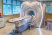
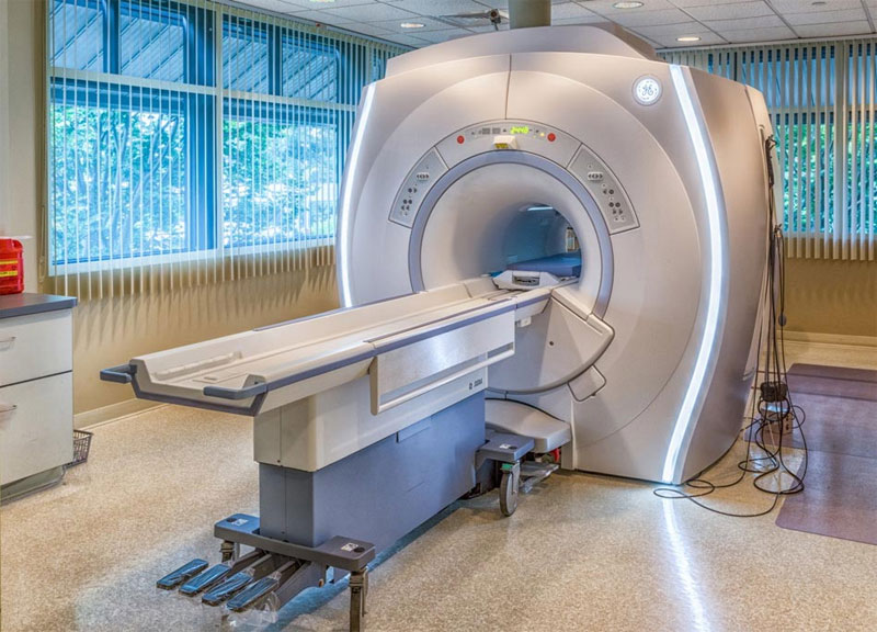
The individual rests within a scanner encircled by a sizable and potent magnet. A magnet designed to receive signals is positioned around the specific area of the body under examination. When capturing images of extensive regions like the abdomen, the receiving magnet is inserted inside the scanner. Conversely, for smaller areas such as a joint, a magnet might encircle the targeted scanning site.
The scanner produces a robust magnetic field that prompts the atoms within the body to align in parallel. Brief bursts of radio waves are emitted from a radiofrequency source, temporarily disrupting the atom alignment. As the atoms return to their alignment, they emit minute signals that are captured by the receiving magnet. These signals’ details are conveyed to a computer, which constructs an image by considering the signals’ intensity and positions.
MRI images can be improved by employing a contrast agent to emphasize specific bodily structures, such as tumors and blood vessels.
Images obtained through MRI closely resemble those generated through CT scanning, yet they provide heightened differentiation between healthy and irregular tissues. MRI proves valuable in examining the brain and spinal cord, the inner intricacies of the eye and ear, internal organs, and blood circulation.
No recognized hazards or adverse effects are associated. The scans avoid employing ionizing radiation and can be conducted multiple times. Nevertheless, it’s important to note that the scanner might disrupt the operation of pacemakers, hearing aids, and other electronic devices.