
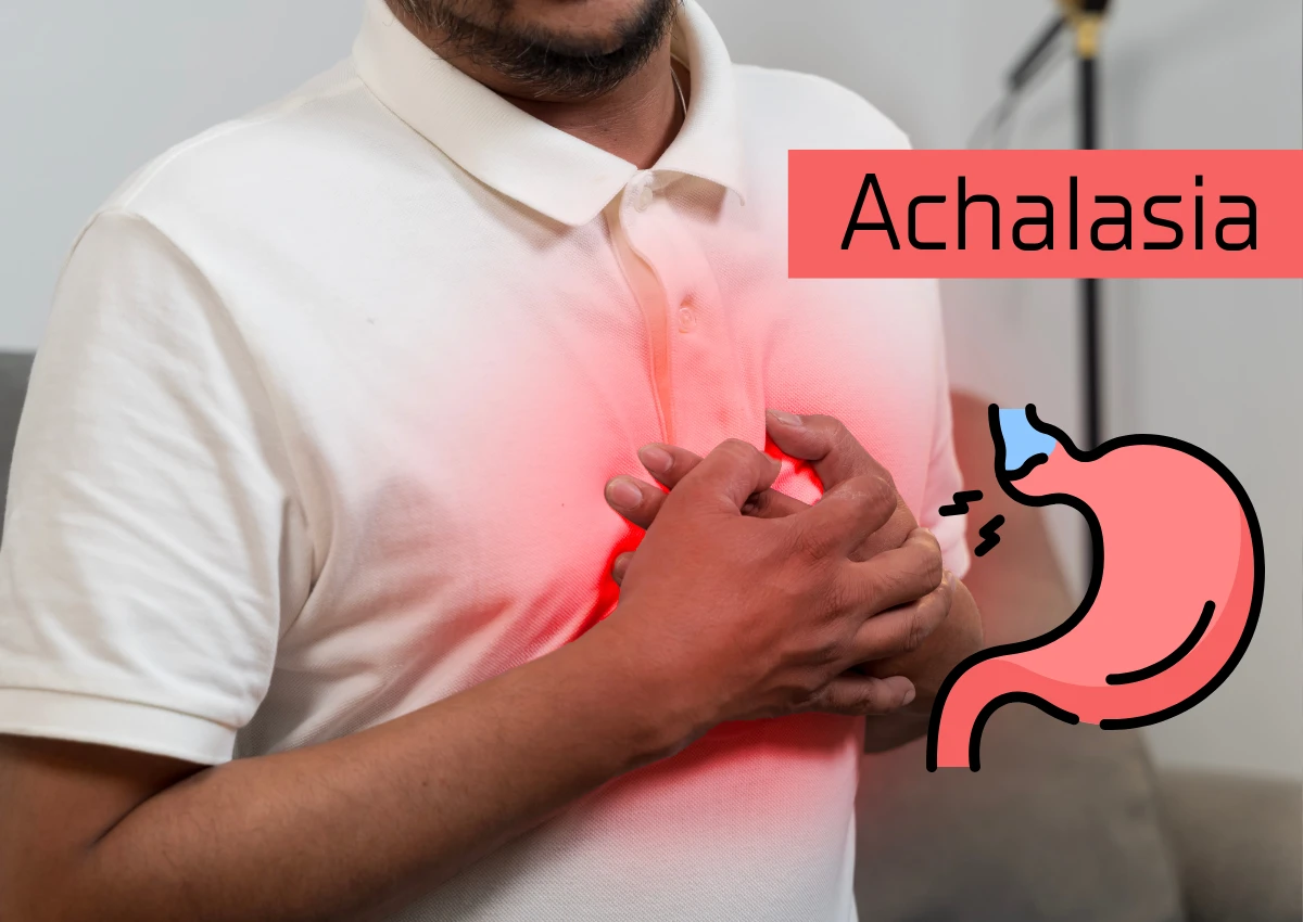
Achalasia is a rare and often misunderstood disorder of the esophagus, the tube that carries food from the mouth to the stomach. In a healthy digestive system, coordinated muscle contractions push food down, and a valve at the bottom—the Lower Esophageal Sphincter (LES)—relaxes to let food enter the stomach. In patients with achalasia, this system fails: the nerves damaging the esophageal muscles stop working correctly, and the LES remains tightly shut, causing food to back up.
For decades, doctors viewed achalasia as a single condition. However, modern diagnostics have revealed that not all cases are the same. Using a gold-standard diagnostic tool called High-Resolution Manometry (HRM), gastroenterologists now categorize the disease into three distinct subtypes using the “Chicago Classification” system. Understanding these types—Type I, II, and III—is critical, as the specific variation dictates not only the symptoms a patient feels but also which treatment will be most effective.
Type I: Classic Achalasia
Type I, often referred to as “Classic Achalasia,” represents a state where the esophageal muscle has effectively ceased to function. On a manometry reading, the esophagus shows aperistalsis, meaning there is zero muscle movement or wave-like contractions (peristalsis) within the esophageal body.
In this scenario, the esophagus acts merely as a passive, gravity-dependent tube. When a patient swallows, there is no muscular push behind the food; it simply sits in the esophagus until the weight of the column of liquid or food forces it through the tight valve at the bottom.
Clinicians often view Type I as the end-stage progression of the disease. Over time, the esophageal muscles may have “burned out” from working against the closed sphincter, leaving the organ dilated and baggy. While the lack of muscle tone sounds severe, patients with Type I generally respond well to standard treatments like the Heller Myotomy (cutting the muscle) or pneumatic dilation, as the primary goal is simply to open the drain at the bottom.
Type II: Compression Achalasia
Type II achalasia is currently the most commonly diagnosed form of the disorder. It is distinguished by a phenomenon known as pan-esophageal pressurization.
Unlike Type I, where the esophagus is quiet and still, the muscles in Type II are still active—but they are confused. Instead of a coordinated wave that pushes food down sequentially, the entire esophagus squeezes simultaneously from top to bottom. This creates a buildup of pressure within the tube, trapping the food between the squeezing muscles and the non-relaxing valve.
Identifying Type II is good news for patients. Studies consistently show that this subtype has the highest success rate for treatment. Because the esophageal muscles still possess tone and the ability to generate pressure, relieving the obstruction at the LES usually restores gravity-aided swallowing very effectively.
Type III: Spastic Achalasia
Type III is the least common and arguably the most challenging form of the condition. It is characterized by spastic contractions.
In Type III, the esophageal nerves misfire, causing the muscles to contract prematurely and violently. These contractions are uncoordinated and fail to push food effectively, but they are powerful. Consequently, patients with Type III often suffer from a distinct set of symptoms. While they experience the difficulty swallowing (dysphagia) seen in other types, they also frequently report severe, crushing chest pain that can mimic a heart attack.
Treating Type III requires a specialized approach. Because the problem involves muscle spasms higher up in the esophagus—not just a tight valve at the bottom—standard treatments that focus only on the LES may fail. These patients often benefit most from a procedure known as POEM (Peroral Endoscopic Myotomy), which allows the surgeon to cut a longer section of the muscle inside the esophagus to relieve the spasms.
Pseudoachalasia: The Mimic
Before a diagnosis of achalasia is confirmed, physicians must rule out “pseudoachalasia.” This refers to conditions that mimic the symptoms and even the manometry readings of achalasia but are caused by something else.
The most serious cause of pseudoachalasia is a tumor, usually at the gastroesophageal junction. A cancer growing in this area can physically compress the LES, making it appear as though the sphincter refuses to relax. Other causes can include Chagas disease (a parasitic infection). This is why an upper endoscopy (EGD) is a mandatory step in diagnosis—to ensure there is no physical blockage or mass before treating the muscles.
Comments
comments