

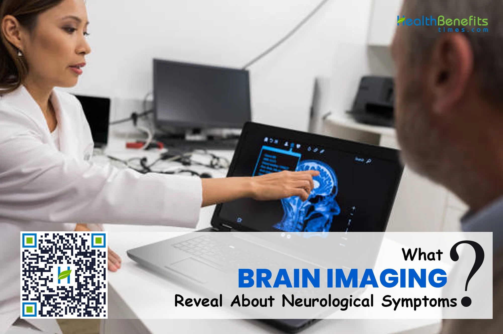
Brain imaging gives doctors a way to look for structural and blood vessel changes that may be connected to neurological symptoms. But a scan is not a simple yes-or-no test.
Different imaging methods answer different questions, and the findings need to be considered alongside symptoms, medical history, and a neurological examination.
What Brain Imaging Can Actually Show
Brain scans can identify conditions such as bleeding, stroke-related changes, tumors, inflammation, injuries, abnormal fluid buildup, and certain blood vessel problems. The right type of scan depends largely on what doctors are trying to confirm or rule out.
CT Scans Are Often Used When Speed Matters
Computed tomography, commonly called CT, uses X-rays to create detailed cross-sectional images of the head and brain. Because the test can usually be completed quickly, it is often useful in emergencies.
A head CT may help doctors look for:
- Bleeding in or around the brain
- Skull fractures after an injury
- Signs of certain types of stroke
- Large masses or tumors
- Abnormal fluid buildup
- Some blood vessel problems
If someone suddenly develops weakness, difficulty speaking, or other possible stroke symptoms, CT imaging may be among the first tests considered because it can quickly show whether bleeding is occurring.
CT does use ionizing radiation, so doctors consider the patient’s situation and whether another imaging method may be more suitable.
MRI Gives a More Detailed View of Brain Tissue
Magnetic resonance imaging, or MRI, uses a strong magnetic field and radio waves instead of X-rays. It can provide highly detailed images of brain tissue and may identify smaller or less obvious abnormalities.
MRI may be used when someone has seizures, persistent weakness, changes in sensation, problems with balance, unusual headaches, or unexplained changes in memory or thinking.
It can also help doctors investigate conditions affecting the brain and nervous system, including multiple sclerosis, tumors, infections, traumatic injuries, and some forms of stroke.
Before an MRI, patients should tell the imaging team about implanted devices, previous operations, metal in the body, pregnancy, kidney problems, or severe claustrophobia. These details can affect how the scan is performed.
Blood Vessel Imaging Answers Different Questions
Sometimes the concern is not mainly the brain tissue but the arteries and veins supplying it.
CT angiography and MR angiography can provide images of blood vessels and may help doctors identify narrowing, blockage, aneurysms, or abnormal vessel structures. In certain cases, cerebral angiography may provide even more detailed information.
This distinction matters because a standard brain scan and a scan designed specifically to examine blood vessels do not provide the same information.
How Symptoms and Scan Findings Work Together
Neurological symptoms do not always point directly to one condition. Dizziness, for example, may come from the brain, inner ear, medications, blood pressure changes, dehydration, or several other causes.
Imaging therefore works best as one part of a wider medical assessment.
Specialist Interpretation Can Add Important Context
Producing the images is only one part of the process. Someone must then interpret what those images show and decide whether the findings may be connected to the patient’s symptoms.
When brain, spine, head, neck, or nervous-system imaging is involved, specialist interpretation may sometimes be needed. A neuroradiologist in abu dhabi may, for example, assess imaging studies involving neurological structures and blood vessels.
The final imaging report is then considered alongside the patient’s examination, symptoms, and medical history. A finding that appears important on a scan may sometimes be unrelated to the problem being investigated, while a smaller change could provide an important clue.
A Normal Scan Does Not Always Explain Everything
One common misunderstanding is that a normal brain scan automatically means there is no neurological problem.
Some conditions do not produce obvious structural changes on routine imaging. Depending on the symptoms, doctors may also use blood tests, electrical tests, cerebrospinal fluid analysis, nerve studies, or other assessments.
That means the useful question after a normal scan is not simply, “Is everything fine?” A better question is, “What has this test ruled out, and what might need to be checked next?”
What Patients Should Ask About Brain Imaging
Knowing why a scan has been ordered can make the process much clearer. It can also help patients understand the results once they are available.
Before the Scan
Useful questions include:
- What problem is the scan trying to identify or rule out?
- Why was CT, MRI, or another imaging method selected?
- Will contrast material be required?
- Are there medical devices, medicines, or previous operations the imaging team should know about?
- What symptoms would require urgent attention while waiting for the scan?
These questions are particularly useful when symptoms have continued for weeks or months, and several possible causes are being considered.
After the Scan
Patients should look beyond whether a report simply says “normal” or “abnormal.”
It may be more useful to ask:
- Did the scan explain my symptoms?
- Were any unrelated findings found?
- Is another type of imaging required?
- Should another specialist review the findings?
- Are additional tests needed?
Medical reports often contain technical language that can sound worrying without proper context. The treating doctor can explain which findings are relevant and what should happen next.
When Neurological Symptoms Need Urgent Attention
Brain imaging can provide valuable information, but emergency symptoms should never be delayed while waiting for a routine scan.
Sudden weakness or numbness, new difficulty speaking, sudden changes in vision, loss of consciousness, a first seizure, or an unusually severe headache can require immediate medical assessment.
For mild or recurring symptoms, doctors may first look at how long they have been happening, whether they are becoming more frequent, what triggers them, and whether other neurological signs are present.
Conclusion
Brain imaging can reveal changes that symptoms alone cannot explain, including bleeding, stroke-related damage, tumors, inflammation, blood vessel problems, and structural abnormalities. CT, MRI, and vascular imaging each provide different kinds of information.
A scan, however, is only one part of the diagnostic process. The clearest answers usually come from combining imaging findings with the person’s symptoms, physical examination, and medical history. Understanding that connection helps patients ask better questions and make more sense of what their scan results actually mean.
Comments
comments